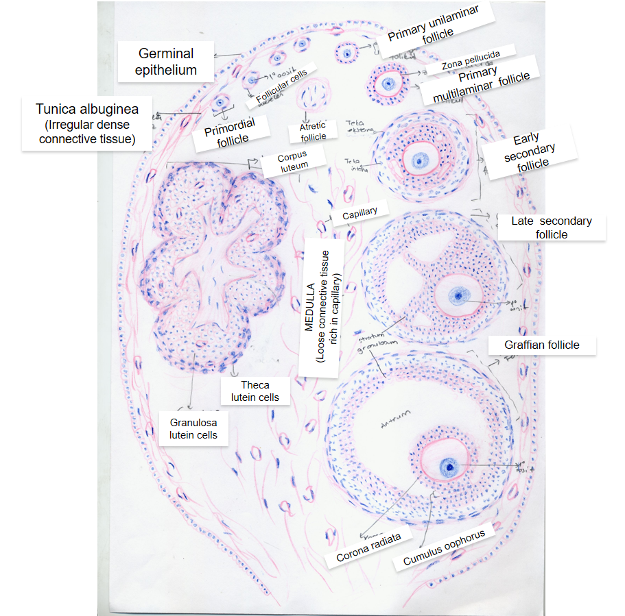
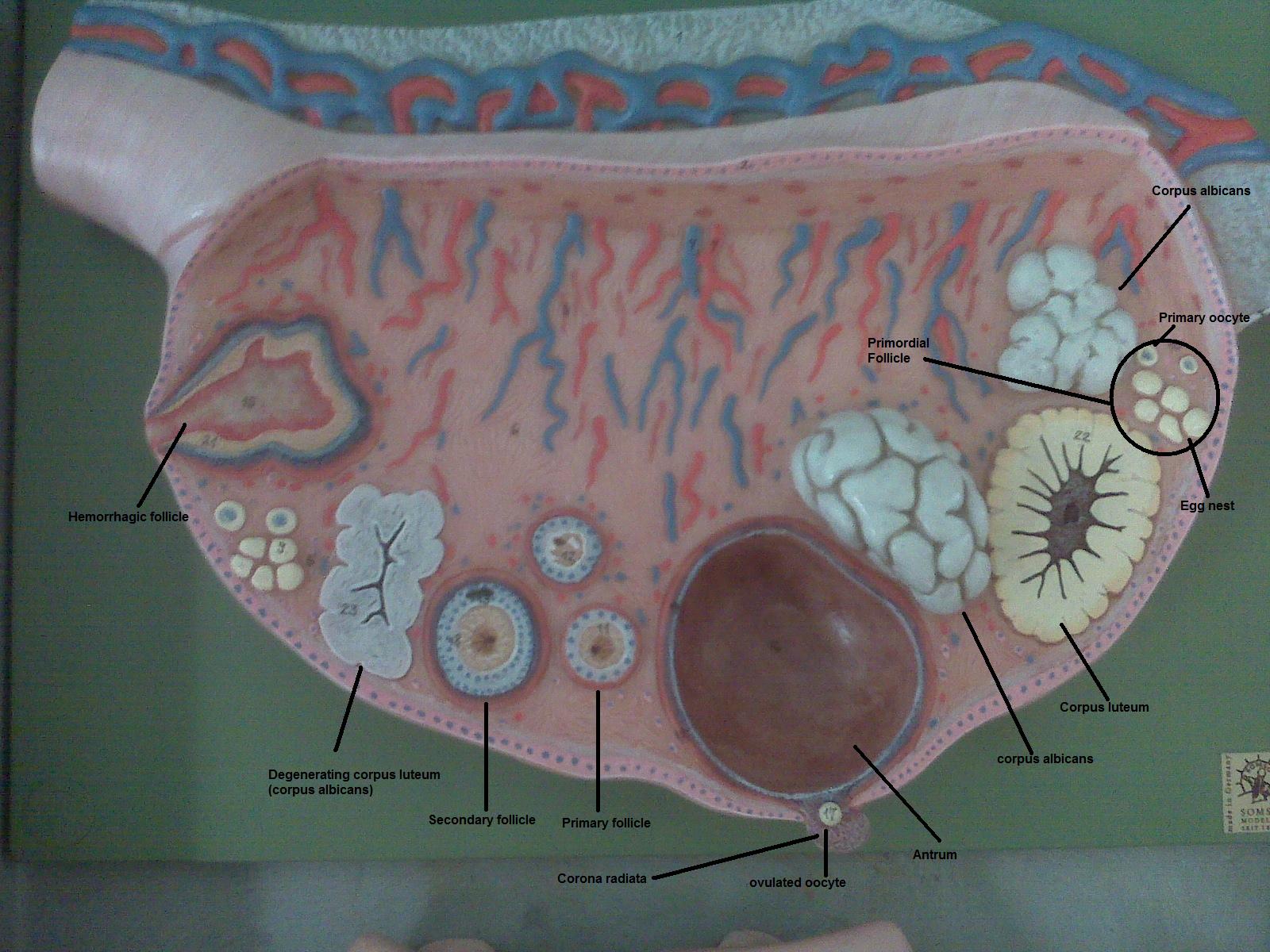
Ovary Histology Diagram
Pin em gene visualisation Ovary histology follicular veterinary ohiostate pressbooks Histology ovary labeled ovarian follicles
Pin em Gene Visualisation
Psc: anatomy and physiology 2: november 2010 Ovary diagrams to print Histology slides database: january 2014
Ovary histology zona pellucida oocyte granulosa ovulation follicle cell secondary file cells cumulus development oogenesis theca embryology developing corpus luteum
The ovary – veterinary histologyOvary histology reproductive female system drawings hematoxylin eosin Ovary reproductive histology ovaries accessmedicine follicle ovarian mhmedical physiology junqueira mcgrawOvary histology general diagrams.
Ovary anatomy labeled model physiology models ovaries corpus luteum psc fertility uterus choose boardFemale reproductive system File:ovary histology 061.jpgIllustrations: ovary.

Follicle histology graafian reproductive biology physiology follicles ovarian primordial microscopic
Ovarian follicals histologyHistology ovary diagram slides resolution high gland .
.

File:Ovary histology 061.jpg - Embryology
PSC: Anatomy and Physiology 2: November 2010

Illustrations: Ovary - General Histology

The ovary – Veterinary Histology

Ovarian Follicals Histology

Ovary Diagrams to Print | 101 Diagrams

Histology Slides Database: January 2014